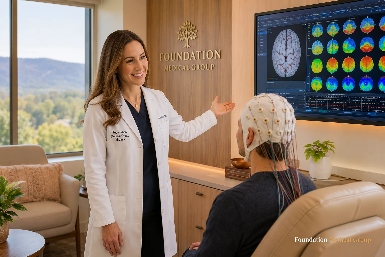
qEEG brain mapping in Virginia is a simple, painless test that reads your brain’s electrical activity and turns it into a picture. Small sensors sit on your scalp, pick up your brainwaves, and a computer compares those signals to typical patterns for your age. The result is a color map that shows how different parts of your brain are working. Doctors use that map to make your care more personal.
If you’re recovering from addiction and feel stuck, this kind of test can add a new layer of insight. It won’t diagnose you, and it won’t cure anything on its own. Think of it as one more helpful tool, sitting next to your doctor, your therapy, and your medicine. Below we’ll explain it in plain words for Virginia readers.
What qEEG Brain Mapping Really Is
Let’s break the name down, because it sounds harder than it is. qEEG stands for quantitative EEG. The “quantitative” part just means a computer measures and counts the brainwaves, instead of a person eyeballing a squiggly line.
Here’s the flow. Sensors read your brain’s activity. The computer sorts those signals by type and location. Then it draws a map that shows where your brainwaves look typical and where they don’t. That map is the “brain mapping” part.
“By type” means by frequency band, and the bands have real numbers behind them: delta below 4 Hz, theta 4 to 7 Hz, alpha 8 to 12 Hz, and beta 13 to 30 Hz. Anything faster than that is usually called gamma. The recording that feeds all of this follows the international 10-20 system, with at least 21 electrodes on an adult scalp including the reference and ground, each checked to sit under 5 kilohms of impedance before the session starts.
You may hear 3 words used together: EEG, qEEG, and brain mapping. They’re closely related, so it’s easy to mix them up. The next section sorts them out.
EEG vs qEEG vs Brain Mapping in Plain Words
These three terms describe steps in one process, not three separate tests. Seeing them side by side makes it click.
| Term | What it means | What it gives you |
|---|---|---|
| EEG | Sensors record your raw brainwaves | A live trace of brain activity |
| qEEG | A computer measures that EEG data | Numbers compared to age norms |
| Brain mapping | Those numbers become a visual | A color map of your brain |
A regular EEG often looks for one thing, like signs of a seizure. qEEG asks a different question. It looks at broad patterns across your whole brain, such as how well areas tied to focus, calm, and mood are working together. So the tool you choose depends on the question your doctor is trying to answer.
Worth knowing what the raw signal is, too. A scalp electrode can’t hear one neuron. It takes roughly 10 square centimetres of cortex firing together to make a visible deflection, so every number in a qEEG report describes a crowd rather than a cell. Modern clinical systems record from 128 channels or more at sampling rates above 10 kHz, which is why the software has enough data to do the counting at all.
How qEEG Personalizes Addiction Recovery
Recovery isn’t one-size-fits-all, and neither is your brain. Substance use can shift the brainwaves linked to impulse control, reward, and calm. A brain map can show some of those shifts in a way that feelings and words alone can’t.
According to the National Institute on Drug Abuse (NIDA), addiction changes how the brain’s circuits work, which is why recovery takes time and support. Seeing a picture of those patterns can help your care team plan smarter. They can pace your therapy, adjust your goals, and coordinate the map with your medicine.
Medication does the structural work in the meantime, and it’s worth keeping the scale in view. In a 2017 BMJ meta-analysis by Sordo and colleagues, all-cause mortality was 4.3 per 1,000 person-years for people on buprenorphine against 9.5 per 1,000 for people off it, with overdose deaths at 1.4 versus 4.6. No brain map moves a number like that. It just helps you shape what sits around the medicine.
This matters most when things overlap. Depression, anxiety, and trouble focusing often ride along with addiction. A map can hint at which of those the brain is struggling with, so your plan targets the right spots. You can read more on the whole approach at our brain mapping in Virginia hub.
Who Might Consider qEEG in Virginia
This test isn’t right for every patient, and that’s fine. Plenty of people do well with standard care and don’t need it. Others feel stuck, and a fresh angle helps.
- You’ve tried standard care and still feel off. Symptoms hang on even though you’re doing the work.
- Several things overlap. Recovery, low mood, worry, and focus troubles all tangle together.
- You’re on Suboxone or buprenorphine and want a clearer view of how your brain is settling.
- You’re curious about neurofeedback, a training method that often uses a brain map as its starting point.
If any of these sound like you, it’s worth a conversation. Your doctor can tell you whether a map would actually add something useful, or whether your current plan is already on track.
What to Expect During a qEEG Session

The test is calmer than most people picture. There are no needles, no shots, and no sedation. You stay awake and comfortable the whole time.
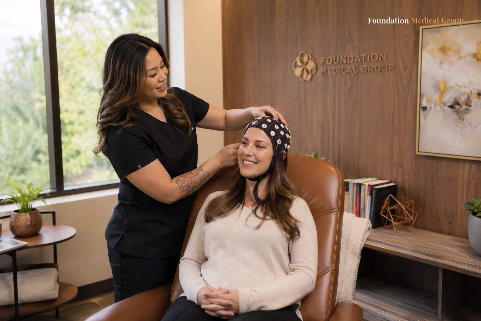
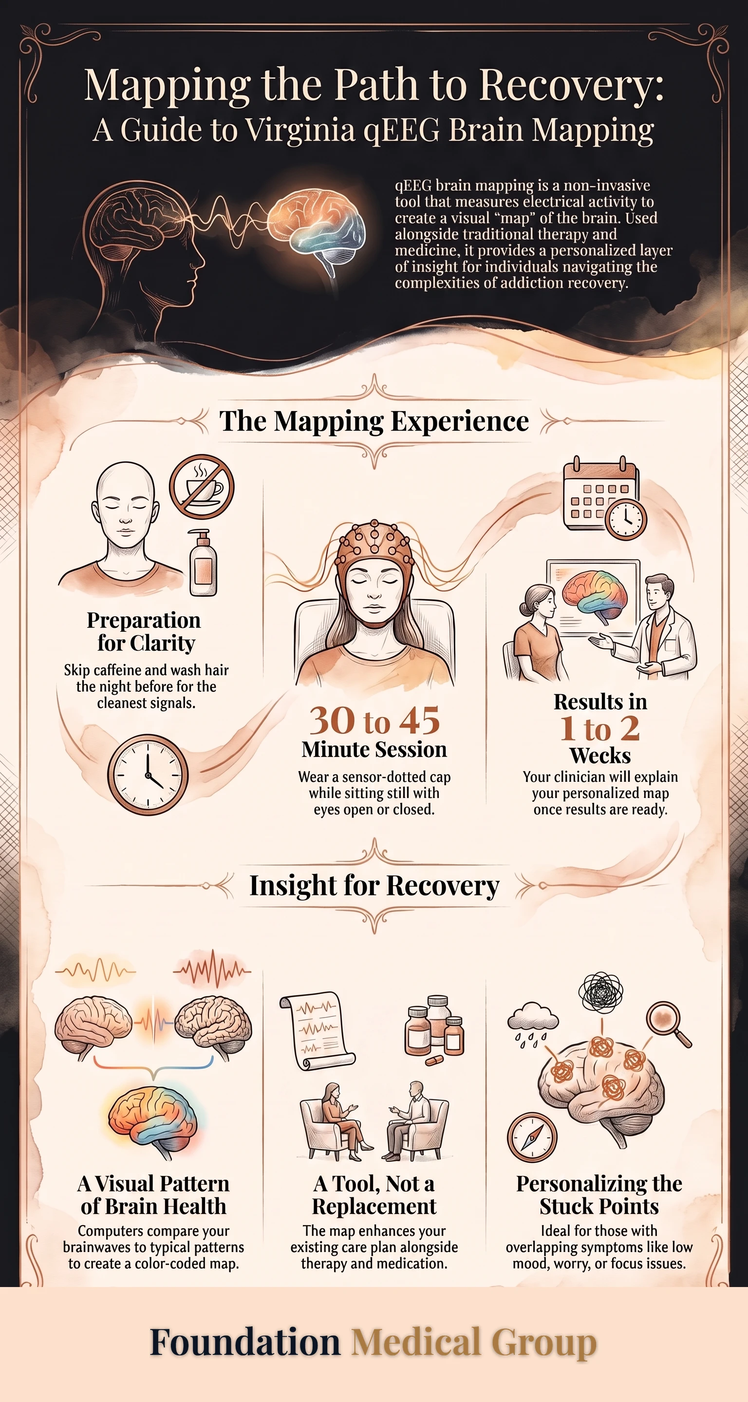
A soft cap dotted with sensors goes on your head. You’ll sit still, sometimes with your eyes open, sometimes closed, and follow a few easy instructions. The eyes-open and eyes-closed segments matter, because alpha activity in the 8 to 12 Hz band changes sharply between the two. Most sessions run about 30 to 45 minutes.
Simple prep helps too. Skip caffeine that day, wash your hair the night before, and leave out conditioner and styling product, since anything left on the scalp pushes electrode impedance past that 5 kilohm target and adds noise to every channel.
Then you wait. Results usually take about one to two weeks to process. When they’re ready, your clinician walks you through the map in plain language and connects it to your next steps.
Honest Talk About Accuracy and Limits
Now the part too many articles skip. qEEG is helpful, but it has real limits, and you deserve the straight story.
First, it can’t diagnose a condition. A map shows activity patterns, not disease labels. Second, the results can wobble. Poor sleep, stress, and certain medicines can all nudge your brainwaves on the day of the test. Which is why a trained provider reads the map with care and treats it as one input, not the whole picture.
Regulators draw the same line. Take the Neuropsychiatric EEG-Based Assessment Aid, or NEBA, which holds an FDA de novo classification under regulation 21 CFR 882.1440. It is narrow by design. It reads one ratio, theta over beta, at one electrode called Cz, in patients aged 6 to 17. The FDA summary states the device is not to be used as a stand-alone and should support a completed clinical evaluation rather than substitute for one. Anyone selling a brain map as an answer by itself is going further than the evidence does.
According to the Substance Abuse and Mental Health Services Administration (SAMHSA), effective care for substance use blends medicine, counseling, and support tailored to each person. A brain map fits inside that blend as a support tool. It’s a helpful lens, not a cure, and it should sit alongside a real medical evaluation, not replace one. If cost is on your mind, our guide on qEEG cost and insurance breaks down what to ask.
How the Results Shape Your Plan
A map only matters if it leads somewhere. Good clinicians turn the picture into clear, doable steps. They line the map up next to your story, your history, and your goals.
From there, a plan might add neurofeedback training, adjust the focus of your therapy, or fine-tune your daily routine. If you’re managing low mood or worry alongside recovery, the map can guide that too. See how the pieces connect on our page about Suboxone with co-occurring depression and anxiety.
The point is coordination. Your medicine, your counseling, and your brain data all pull in the same direction, toward steady, long-term progress.
Infographic: The qEEG Brain Mapping Journey in Virginia
Frequently Asked Questions
What is qEEG brain mapping used for?
It’s used to picture how your brain is working so care can be more personal. Clinicians read the map to guide therapy, plan neurofeedback, and coordinate care with your recovery. It supports decisions; it doesn’t make them alone.
Is qEEG brain mapping safe?
Yes. The test only listens to your natural brainwaves through sensors on your scalp. Nothing is sent into your brain, there are no needles, and there’s no sedation.
Does qEEG diagnose addiction, depression, or ADHD?
No. A brain map can’t diagnose any condition. It shows activity patterns that your doctor reads alongside your history and symptoms to build a fuller picture.
Can I get qEEG while I’m on Suboxone or buprenorphine?
Yes. Brain mapping can run right alongside your medication without changing it. Many people use it to better understand how their brain is settling during recovery.
How long does a qEEG session take in Virginia?
The test itself usually lasts about 30 to 45 minutes, and you stay awake and comfortable. Your results are typically ready in about one to two weeks, when your clinician explains them.
Key Takeaways and Next Steps
Here’s what to hold on to. qEEG brain mapping in Virginia turns your brainwaves into a picture, so your care can fit you instead of the average. It won’t diagnose or cure, but it can add real insight, especially when recovery, mood, and focus all overlap. Used well, it sits next to your doctor, your therapy, and your medicine.
- Remember the plain version: sensors read your brainwaves, a computer measures them, and a map shows the patterns.
- Treat the map as a helpful tool, not a final answer or a replacement for a medical exam.
- Ask your provider whether a map would add something useful to your current plan.
- If several things overlap in your recovery, mention it, since that’s where a map often helps most.
Ready to learn more? Reach out to Foundation Medical Group and ask whether qEEG brain mapping fits your recovery. One honest conversation can turn a confusing idea into a clear, personal next step.
Sources
- Electroencephalogram, StatPearls, NCBI Bookshelf.
- Normal EEG Waveforms, StatPearls, NCBI Bookshelf.
- FDA de novo decision summary K112711, Neuropsychiatric EEG-Based Assessment Aid (NEBA).
- Sordo et al., Mortality risk during and after opioid substitution treatment, BMJ 2017.
- Substance Abuse and Mental Health Services Administration (SAMHSA), Find Treatment.